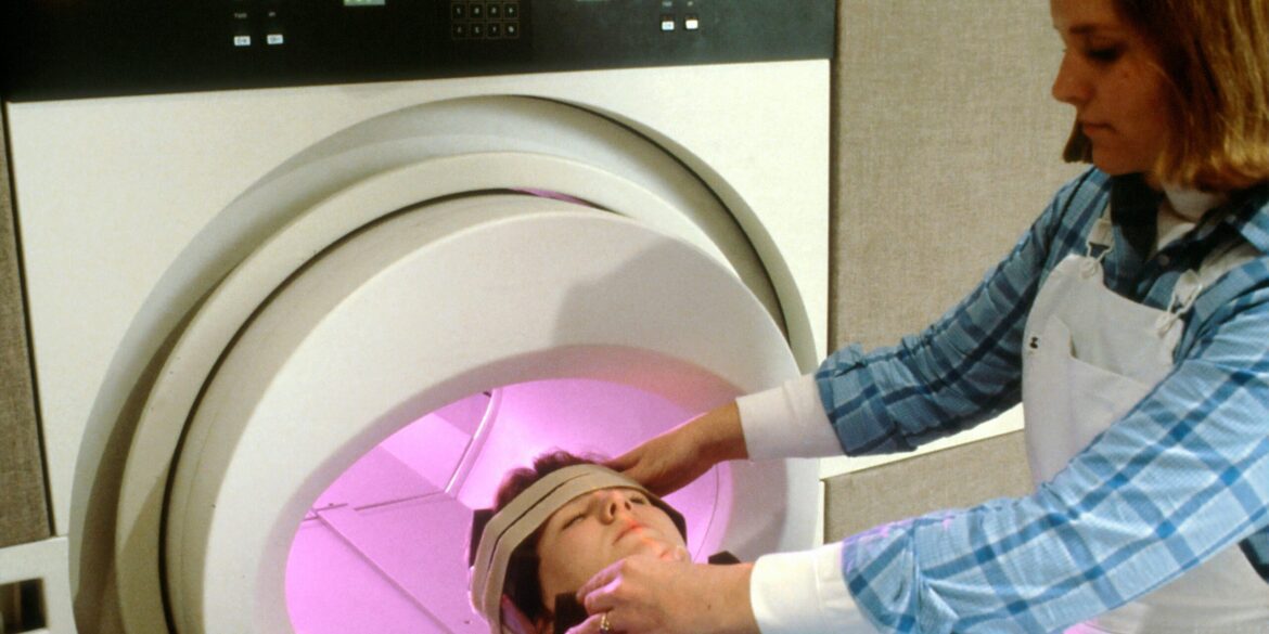
A research team supported by the U.S. National Institutes of Health (NIH) has unveiled a groundbreaking MRI scanner, dubbed Connectome 2.0, capable of noninvasively imaging microscopic nerve structures within the living human brain with near‑cellular resolution. Detailed results were published on July 16, 2025, in Nature Biomedical Engineering.
Connectome 2.0 represents a leap forward from standard MRI systems by combining two technical innovations: a head‑conforming design, which snugly surrounds the participant’s head to enhance spatial precision, and a significantly increased number of signal‑channel interfaces that dramatically improve signal‑to‑noise ratio. These advancements allow the system to visualize nerve fibers and cell‑like structures such as axon diameters and individual cell sizes down to single‑micron precision in living volunteers.
Previously, imaging at such scale was only possible postmortem or in animal studies. Early reports confirm the system is safe for use in healthy research subjects and already able to detect subtle microstructural differences across individuals.
The project is part of NIH’s broader BRAIN CONNECTS initiative, a segment of the larger BRAIN Initiative, which aims to map the wiring diagram—or “connectome”—of the human brain across multiple scales. John Ngai, Director of NIH’s BRAIN Initiative, described the innovation as a transformative leap in brain imaging, pushing the boundaries of what we can see and understand about the living human brain at a cellular level.
Read Also: https://empirestatereview.com/breakthrough-gene-therapy-offers-new-hope-for-congenital-deafness/
Senior author Susie Huang of Massachusetts General Hospital’s Department of Radiology said the goal was to build an imaging platform that could truly span scales from cells to circuits. She noted that the new system provides researchers and clinicians with a powerful tool to study brain architecture in health and disease in real time.
Experts highlight several areas where this technology could have immediate impact. These include early diagnosis and monitoring of neurological and neuropsychiatric conditions such as Alzheimer’s, Parkinson’s, schizophrenia, developmental disorders, and epilepsy, by spotting microstructural disruption before symptoms emerge. It also supports precision neurology, where individualized brain profiles can guide therapies and targeted brain stimulation strategies based on a person’s unique neural wiring. Additionally, the system helps link cell-level abnormalities with broader network dysfunctions to better understand cognition, behavior, and disease progression.
Connectome 2.0 builds on NIH’s long-standing investment in in vivo MRI hardware and methodology, particularly in ultra‑high field imaging, novel gradient coil design, and advanced reconstruction algorithms aimed at improving speed and spatial resolution.
The project’s publication in Nature Biomedical Engineering underscores an interdisciplinary partnership across NIH and external institutions through the BRAIN Initiative framework. The scanner’s design reflects years of technological advancement, including previous Connectome 1.0 systems and developments in diffusion‑based tractography and microstructural modeling.
The introduction of Connectome 2.0 may redefine how researchers and clinicians approach brain disorders. Noninvasive cellular resolution allows repeated imaging in individuals, opening possibilities for longitudinal tracking of disease progression or treatment response. The technology advances the possibility of integrating real-time imaging with stimulation therapies, potentially enabling next‑generation, patient‑specific neuromodulation. As trials and refinements continue, this scanner could influence future clinical tools in major neurology centers, transitioning precision neurology from research to clinical practice.
The NIH‑unveiled Connectome 2.0 MRI scanner marks a landmark achievement in neuroimaging. For the first time, researchers can visualize microscopic nerve structures in living human brains with clarity and resolution previously limited to postmortem or animal studies. Enabled by innovations in coil design, signal channels, and head-conforming architecture, Connectome 2.0 paves the way for precision neurology, enhanced disease understanding, and tailored treatments.
Developed under the BRAIN CONNECTS and BRAIN Initiative programs, the system reflects a commitment to building a comprehensive wiring diagram of the human brain across scales and individuals. As it becomes more widely adopted, Connectome 2.0 holds real promise for diagnosing and treating neurodegenerative and developmental disorders in ways never before possible.

